Thermal Imaging
Don’t just detect breast cancer, prevent it!
Don’t just detect breast cancer, prevent it!
Thermography provides a safe, simple, and painless way to identify developing breast cancer. By the time mammography detects breast cancer, a women has had it for 8 to 10 years. By carefully examining aspects of temperature and blood vessel activity of the breasts in thermal images, signs of possible cancer or pre-cancerous cell growth may be detected within the first year of development. This provides the earliest detection of breast cancer possible.
The digitized data from the camera is saved by the imaging system computer for evaluation by a licensed physician who is trained and certified as a Clinical Thermologist which allows for the specific interpretation of your thermal patterns.
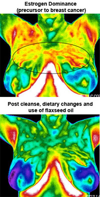 By the time mammography detects breast cancer, a women has had it for 8 to 10 years. By carefully examining aspects of temperature and blood vessel activity of the breasts in thermal images, signs of possible cancer or pre-cancerous cell growth may be detected within the first year of development. This provides the earliest detection of breast cancer possible. Because of thermal imaging’s extreme sensitivity, these temperature variations and vascular changes are among the earliest signs of breast cancer or a pre-cancerous state of the breast.
This breast imaging procedure is based on the principle that chemical and blood vessel activity in both pre-cancerous tissue and the area surrounding a developing breast tumor is almost always higher than in the normal breast. Since pre-cancerous and cancerous masses are highly metabolic tissues, they need an abundant supply of nutrients to maintain their growth. To obtain these nutrients they increase circulation to their cells by secreting chemicals to keep existing blood vessels open, recruit dormant vessels, and create new ones (neoangiogenesis). This process results in an increase in regional surface temperatures of the breast.
State-of-the-art applications use ultra-sensitive thermal imaging cameras and sophisticated computer software to detect, analyze, and produce high-resolution diagnostic thermal images of these temperature and vascular changes.
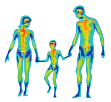
Thermography allows multiple views of the body from a physiological and functional point of view. This provides views of your body as a whole for diagnostic insight and a perspective of individual wellness without compromising any systems (100% safe).
Your body is considered a thermodynamic living organism that forwards and receives information by way of the nervous system which is medically divided into three (3) primary systems: Motor = muscle control, Sensory = touch and vision, and Autonomic = control of blood flow to all organs.
Skin is your largest organ and has unique and distinctive thermal patterns that are specific to you. Skin thermal patterns demonstrate hot and cold areas that are consistent with the body part involved in that region and are imaged with highly sensitive medical infrared cameras.
The process begins with simple preparation steps that are listed below (patient preparation instructions) that must be strictly followed before you arrive. Once in the office, you will be asked to complete a questionnaire on your breast health history. You will then meet with our thermographic technician to go over the procedure and review your questionnaire. During the examination you will be disrobed from the waist up for both imaging and to allow for the surface temperature of the body to acclimate with the room temperature. A female technician will perform the imaging for you. There will be a visual inspection of the surface of the breasts so that we can correlate any surface findings with the infrared images. Once this is done, you will be left for 15 minutes to allow your body to acclimate with the special temperature conditions of the room. After this time, you will be positioned in front of the infrared camera so that all of the surfaces of the breasts, upper chest, and under arms are imaged.
The images are captured by a high resolution digital infrared imaging camera and sent to a computer software program for storage and analysis (the images are archived for comparison of future images so that the breasts can be monitored over time). Sophisticated computer programs allow the doctor to isolate temperature differentials, perform analyses, and create a thorough report.
Once the images have been taken, our Board Certified Clinical Thermologist will digitally process and grade the images, and a full written report (including your images) will be mailed to you within 10 days to 2 weeks.
The following instructions must be strictly adhered to before you arrive for your appointment:
Note: Breast implants, breast reductions, and biopsies do not interfere with infrared imaging. Breast thermography is safe to have during pregnancy or when nursing. The procedure may also be performed during any part of the menstrual cycle without effecting the interpretation of the images.